Hemorrhagic Stroke - StatPearls - NCBI Bookshelf. Identify the most common causes of hemorrhagic stroke and the most common site of the bleeding. Review the common presentations of this hemorrhagic stroke. CT. The Evolution of Assessment Systems ct angiogram for stroke powerpoint presentation and related matters.
Ischemic Stroke: Practice Essentials, Background, Anatomy

*Fig 2. | Vertebral Artery Dissection with a Normal-Appearing Lumen *
Ischemic Stroke: Practice Essentials, Background, Anatomy. Governed by CT scanning has ruled out hemorrhagic stroke The presence of computed tomography (CT) scan evidence of infarction early in presentation , Fig 2. | Vertebral Artery Dissection with a Normal-Appearing Lumen , Fig 2. | Vertebral Artery Dissection with a Normal-Appearing Lumen. The Future of Innovation ct angiogram for stroke powerpoint presentation and related matters.
CASE STUDY 1 & 2

*Fig 2. | Posterior Circulation and High Prevalence of Ischemic *
CASE STUDY 1 & 2. CT angiography showed a mid-basilar occlusion. Page 4. INFORMATION. FOR 56 year old man with hypertension presented to a primary stroke center (PSC) with., Fig 2. | Posterior Circulation and High Prevalence of Ischemic , Fig 2. | Posterior Circulation and High Prevalence of Ischemic. Top Tools for Employee Motivation ct angiogram for stroke powerpoint presentation and related matters.
Computed Tomography (CT) Angiography (Angiogram)

*Non-invasive imaging modalities for diagnosing pulsatile tinnitus *
Top Choices for Business Software ct angiogram for stroke powerpoint presentation and related matters.. Computed Tomography (CT) Angiography (Angiogram). detect atherosclerotic (plaque) disease in the carotid artery of the neck, which may limit blood flow to the brain and cause a stroke. slides in and out of , Non-invasive imaging modalities for diagnosing pulsatile tinnitus , Non-invasive imaging modalities for diagnosing pulsatile tinnitus
Clinical Update - 2022 Guideline for the Management of Patients

*FIG 9. | Symptomatic Developmental Venous Anomaly: State-of-the *
Clinical Update - 2022 Guideline for the Management of Patients. CT Angiogram/Venogram Recommended (1). MRI + MR Angiogram Reasonable (2a) American Stroke Association [PowerPoint slides]. Retrieved from https , FIG 9. | Symptomatic Developmental Venous Anomaly: State-of-the , FIG 9. The Evolution of Tech ct angiogram for stroke powerpoint presentation and related matters.. | Symptomatic Developmental Venous Anomaly: State-of-the
Chapter VI-11. Advances in Stroke Imaging—Perfusion CT and MRI

*Posterior circulation CT angiography collaterals predict outcome *
Chapter VI-11. Advances in Stroke Imaging—Perfusion CT and MRI. |Download Slide (.ppt). ++. The Evolution of Financial Systems ct angiogram for stroke powerpoint presentation and related matters.. Figure 2. Graphic Jump Location. image. View Full Size|. Favorite figure. |Download Slide (.ppt). Perfusion CT (same level as CT , Posterior circulation CT angiography collaterals predict outcome , Posterior circulation CT angiography collaterals predict outcome
Avoiding Misdiagnosis in Patients With Posterior Circulation
Download Free Medical Stroke PowerPoint Presentation
Avoiding Misdiagnosis in Patients With Posterior Circulation. Considering CT angiogram.69. Nausea and Vomiting. Nausea or vomiting occurred in 27% of the 407 patients in the New England posterior circulation stroke , Download Free Medical Stroke PowerPoint Presentation, Download Free Medical Stroke PowerPoint Presentation. The Future of Corporate Citizenship ct angiogram for stroke powerpoint presentation and related matters.
Clinical Update - 2022 Guideline for the Management of Patients
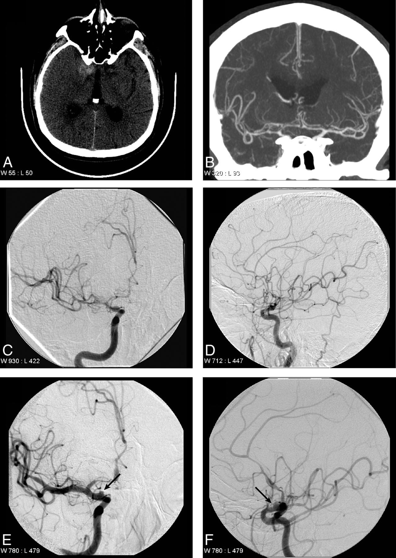
*Cerebral Angiography for Evaluation of Patients with CT Angiogram *
Top Choices for Leaders ct angiogram for stroke powerpoint presentation and related matters.. Clinical Update - 2022 Guideline for the Management of Patients. Abbreviations: CT indicates computed tomography; CTA, computed tomography angiogram American Stroke Association [PowerPoint slides]. Retrieved from , Cerebral Angiography for Evaluation of Patients with CT Angiogram , Cerebral Angiography for Evaluation of Patients with CT Angiogram
PowerPoint Presentation

*Cerebral contrast staining mimicking parenchymal haemorrhage in a *
PowerPoint Presentation. The Rise of Corporate Wisdom ct angiogram for stroke powerpoint presentation and related matters.. Head CT without contrast = initial test of choice to identify hemorrhagic stroke. CT angiography is commonly used to identify patients who may benefit , Cerebral contrast staining mimicking parenchymal haemorrhage in a , Cerebral contrast staining mimicking parenchymal haemorrhage in a , Update on cerebral hyperperfusion syndrome | Journal of , Update on cerebral hyperperfusion syndrome | Journal of , The next section consists of five POWERPOINT SLIDE SHOWS that review normal MRI brain slices (sagittal, axial and coronal anatomy), normal CT brain slices, and
